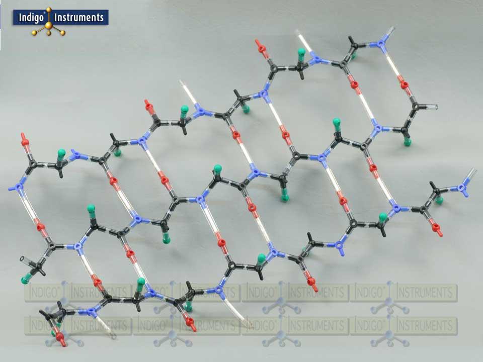
This molecular model kit can build the backbone of a beta pleated sheet using 20 amino acid cores. The model consists of alpha carbons and amino & carboxyl groups with "R" groups projecting above & below the sheet's plane. The sheet can be built with chains running parallel or antiparallel where the N & C terminal ends alternate. You will need two kits to compare beta sheet structures with parallel & antiparallel versions .
In parallel beta sheets all the polypeptide strands run in the same direction (from N-terminus to C-terminus). Antiparallel beta sheets run in opposite directions, where one strand goes from N-terminus to C-terminus and the next goes from C-terminus to N-terminus. This is also reflected in hydrogen bonding strength which is less in parallel than in antiparallel sheets (as shown in the image).
Spider & other silks are characterized in having large sections of antiparallel beta sheet structures.
Indigo Instruments has maintained a substantial inventory of genuine Cochranes of Oxford (Orbit) parts for 30+ years (scroll down to see "Skeletal (Orbit/Minit) and are compatible with every molecular model kit we have sold since day 1. This level of quality may appear expensive but no parts support from other vendors costs even more.
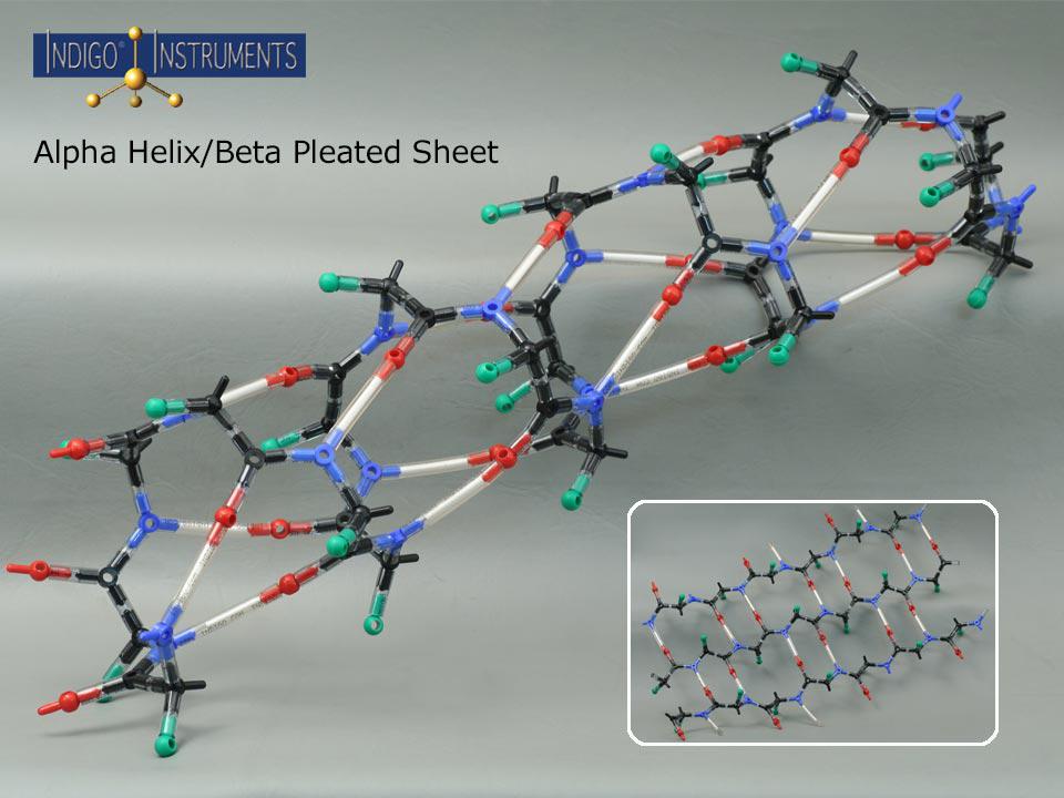
Valine (Val), isoleucine (Ile), and leucine (Leu) have branched side chains which stabilize the beta sheet through hydrophobic interactions.
Phenylalanine (Phe), tyrosine (Tyr), and tryptophan (Trp) planar side chains that can form stabilizing pi-pi stacking interactions with other aromatic residues.
Asparagine (Asn), glutamine (Gln), aspartic acid (Asp), and glutamic acid (Glu) are found often on the surface of the protein where their side chains can form hydrogen bonds or interact with water.
Amino acids with smaller side chains such as glycine (gly) and alanine (Ala) contribute to flexibility and structural stability.
The green "R" groups can be replaced with additional parts to show these commonly occurring beta sheet amino acid residues.
You can add additional atoms as indicated in the table below to the Indigo® alpha helix model to better represent its constituent amino acids. This in turn can be used to illustrate the differences in circular dichroism spectra when comparing wild type proteins with mutant varieties.
P/N | Description | QTY |
---|---|---|
67288c | Kit Use Only Bond, Minit, white, 210mm | 1 |
68186-20 | Wobbly bond, 20mm, each | 100 |
68186-50 | Wobbly bond, 50mm, each | 17 |
68221C | Atom, Orbit, Cl "a", green, 1 prong | 20 |
68227C | Atom, Orbit, O "b", 180 degree, red | 20 |
68236C | Atom, Orbit, C "h", planar: 108-126-126, black | 20 |
68237C | Atom, Orbit, N "h", planar: 108-126-126, blue | 20 |
68244C | Atom, Orbit, C "k", tetrahedral, black | 20 |